The Role of Albumin in the Anion Gap
Albumin is the single largest contributor to the normal anion gap — and the reason a critically ill patient's raw anion gap can look deceptively normal in the face of a serious organic acidosis. Here is the physiology and the correction rule that fixes it.
Albumin: the dominant "unmeasured" anion
When clinicians call the anion gap a measure of unmeasured anions, the largest of those unmeasured anions is hiding in plain sight on every metabolic panel: albumin. Human serum albumin is a 66 kDa protein with a physiological isoelectric point below 5, so at plasma pH of 7.40 it carries a substantial net negative charge — roughly 0.6–0.7 mEq of negative charge per gram per decilitre, contributed mainly by its abundant aspartate and glutamate residues1. At a normal serum albumin of ~4.0 g/dL, albumin alone accounts for approximately 2.5 mEq/L per g/dL × 4 ≈ 7–8 mEq/L, or about 75% of the entire normal anion gap12.
This makes albumin the dominant force setting the "normal" gap. Phosphate and sulfate together contribute only ~2 mEq/L; organic anions (lactate, ketones) contribute ~1–2 mEq/L in health. Pull albumin out and the floor of the normal range drops with it. This is the entire basis for understanding the unmeasured anions composition and for why a low albumin — extremely common in the ICU, in cirrhosis, in nephrotic syndrome, and in malnutrition — silently lowers the apparent anion gap.
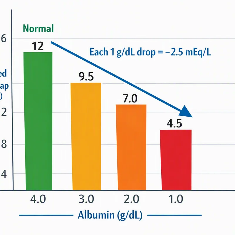
For every 1 g/dL the serum albumin falls below 4.0 g/dL, the expected normal anion gap drops by approximately 2.5 mEq/L. A patient with albumin of 2.0 g/dL therefore has a new "normal" gap of roughly 12 − (2 × 2.5) = 7 mEq/L rather than 12. A measured gap of 10 mEq/L — which looks normal — is in fact markedly elevated for that patient.
The albumin-corrected anion gap formula
The Figge equation restores comparability between hypoalbuminemic and normoalbuminemic patients by adding back the charge that albumin should be carrying:
Worked example: a septic ICU patient has Na⁺ 138, Cl⁻ 108, HCO₃⁻ 20, albumin 1.8 g/dL. The raw gap is 138 − (108 + 20) = 10 mEq/L, which sits in the textbook "normal" range of 8–12. But applying the corrected anion gap formula: 10 + 2.5 × (4.0 − 1.8) = 10 + 5.5 = 15.5 mEq/L. The corrected gap reveals a high-anion-gap metabolic acidosis that the raw number completely hid. You can run this exact scenario in the corrected anion gap calculator and read the deeper rationale on why albumin matters in interpretation.
Why every critically ill patient needs correction
Hypoalbuminemia is essentially universal in the critically ill. Sepsis drives capillary leak and redistribution; hepatic dysfunction reduces synthesis; malnutrition and inflammation lower baseline levels. ICUs routinely see albumin between 1.5 and 2.5 g/dL, which means the conventional "normal" 8–12 mEq/L range overstates the true normal gap for these patients by 4–6 mEq/L. The practical consequence is well documented: an uncorrected anion gap misses roughly half of clinically significant high-anion-gap metabolic acidoses in hypoalbuminemic patients.
This is the reason albumin correction is now standard teaching rather than a research curiosity. The electrolyte panel already contains the albumin value (or it is available on the same draw as the comprehensive metabolic panel), so there is no additional cost or delay. The only failure mode is forgetting to apply the correction — which is why every anion gap reported in a critically ill patient should be reported as a corrected value, and why our calculator applies the Figge adjustment automatically when an albumin is entered.
Hyperalbuminemia is rare, but acute dehydration can push albumin above 5.0 g/dL and transiently raise the uncorrected gap. In that setting the corrected value will pull the apparent gap down, occasionally normalizing a borderline-high reading. Always interpret the corrected gap in the full clinical context, not in isolation.
A note on the other weak acids
Albumin is the dominant non-volatile weak acid in plasma (the "A⁻" in the Henderson-Hasselbalch total weak-acid term), but it is not the only one. Globulins and inorganic phosphate contribute the remainder. In multiple myeloma, cationic (positively charged) paraproteins can invert the relationship entirely, producing a pathologically low anion gap — a rare but classic board clue. For the vast majority of patients, however, albumin is the only weak acid whose concentration varies enough to matter for day-to-day anion gap interpretation, which is why the Figge equation uses albumin alone.
Reference: Figge J, et al. "The role of serum proteins in acid-base equilibria" (Crit Care Med)1; Kraut & Madias, StatPearls "Anion Gap"2; the LITFL albumin-correction page3. The 2.5 mEq/L per 1 g/dL coefficient is the consensus value used across these sources.
References
- Figge J, et al. "Anion gap and hypoalbuminemia." Critical Care Medicine. 1998; 26(11):1807–1810. Link ↗
- Kraut JA, Madias NE. "Serum anion gap: uses and limitations." Clinical Journal of the American Society of Nephrology (CJASN). 2007. Link ↗
- LITFL. "Albumin-Corrected Anion Gap." Life in the Fast Lane. Link ↗
- Figge J, et al. "The role of serum proteins in acid-base equilibria." Critical Care Medicine. 1991. Link ↗