Mixed Acid-Base Disorders & the Delta-Delta
When two acid-base disturbances occur at once, no single rule of compensation can fit the numbers. The delta-delta method compares the rise in anion gap to the fall in bicarbonate and exposes the second disorder hiding inside the first.
What is a mixed acid-base disorder?
A mixed acid-base disorder is the simultaneous presence of two or more primary acid-base disturbances in the same patient. A pure disorder follows predictable compensation rules — Winter's formula for metabolic acidosis, the Boston rules for respiratory disorders. When the measured compensation falls outside the predicted range, or when the anion gap and bicarbonate do not move in lockstep, a second primary process is at work.
Mixed disorders are not rare. They are the rule in the critically ill, where multiple insults — vomiting, diarrhea, shock, diuretics, mechanical ventilation — stack on top of one another. Recognizing them matters because each primary process has its own cause and its own treatment. Treating only one leaves the other to drive pH in the opposite direction.
The two most common mixed metabolic patterns the anion gap uncovers are a mixed metabolic acidosis (a HAGMA running alongside a hyperchloremic NAGMA — for example, sepsis with lactic acidosis plus massive saline resuscitation) and a HAGMA with concurrent metabolic alkalosis (for example, lactic acidosis in a vomiting patient). The delta-delta approach detects both.
The delta-delta method, step by step
The delta-delta (also written Δ-Δ) rests on one physiologic principle: in a pure HAGMA, every molecule of unmeasured acid that widens the anion gap consumes exactly one molecule of bicarbonate. So the rise in anion gap (ΔAG) should equal the fall in bicarbonate (ΔHCO₃⁻). When they do not match, a second process is hidden in the mismatch.
- Calculate the anion gap.
AG = Na⁺ − (Cl⁻ + HCO₃⁻). Use the patient's actual values. - Find the change in anion gap (ΔAG). Subtract the normal midpoint of 12:
ΔAG = AG − 12. - Find the change in bicarbonate (ΔHCO₃⁻). Subtract the observed HCO₃⁻ from the normal midpoint of 24:
ΔHCO₃⁻ = 24 − HCO₃⁻. - Compute the delta ratio.
ΔAG / ΔHCO₃⁻. The delta ratio calculator does this directly; the related delta gap calculator reports the difference(ΔAG) − (ΔHCO₃⁻)in mEq/L. - Interpret the ratio using the bands below.
Delta ratio interpretation bands
The ratio tells you how the anion gap and bicarbonate are moving relative to each other. Each band maps to a specific combination of disorders:
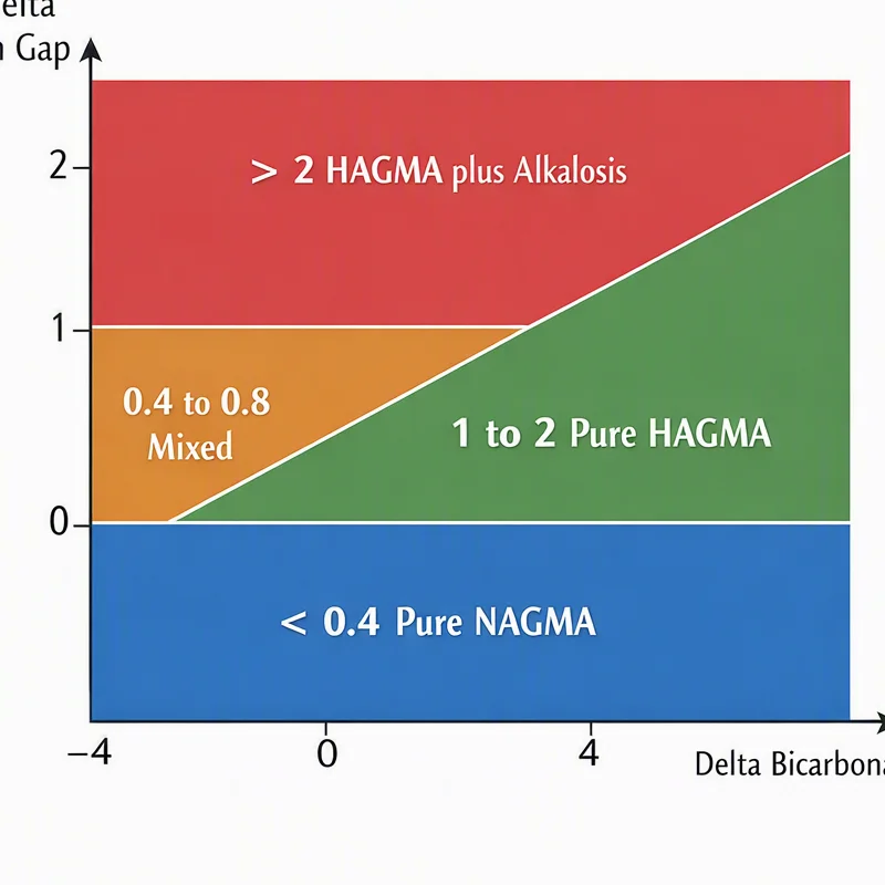
| Delta ratio | Interpretation | What it means |
|---|---|---|
| < 0.4 | Pure hyperchloremic (normal gap) metabolic acidosis | Bicarbonate falls with no rise in gap — classic NAGMA. |
| 0.4 – 0.8 | Mixed HAGMA + NAGMA | Gap rises but less than the bicarbonate falls — a hyperchloremic component is added on top of HAGMA. |
| 1 – 2 | Pure HAGMA (typical ≈ 1–1.6) | Gap and bicarbonate move one-for-one — a single high-anion-gap process. |
| > 2 | HAGMA + metabolic alkalosis | Bicarbonate falls less than expected (or rises) — a concurrent alkali-generating process is offsetting the acid. |
The band between 0.8 and 1.0 is a transitional zone — usually read as "predominantly HAGMA with a possible small hyperchloremic component." No single cut-off is absolute; the bands are heuristics to be interpreted with the clinical context12.
If you prefer absolute numbers to ratios, the delta gap = (AG − 12) − (24 − HCO₃⁻). A delta gap around 0 mEq/L means pure HAGMA; a negative value (e.g. −6) signals concurrent NAGMA; a strongly positive value (e.g. +10) signals concurrent metabolic alkalosis. It is the same information as the delta ratio, just reported as a difference.
Worked example — sepsis with saline resuscitation
A septic patient resuscitated with 4 litres of normal saline has the following blood gas and panel: Na⁺ 140, Cl⁻ 110, HCO₃⁻ 16, albumin 3.8 g/dL.
Step 1 — anion gap: 140 − (110 + 16) = 14 mEq/L. Already above 12, so there is a HAGMA component.
Step 2 — ΔAG: 14 − 12 = +2.
Step 3 — ΔHCO₃⁻: 24 − 16 = +8.
Step 4 — delta ratio: 2 / 8 = 0.25.
Step 5 — interpretation: A ratio of 0.25 sits firmly in the pure hyperchloremic band, even though the anion gap is mildly elevated. The bicarbonate fell by 8 mEq/L but the gap rose by only 2 — the remaining 6 mEq/L of acid load must be coming from a hyperchloremic (saline-induced) source. This is a mixed metabolic acidosis: early lactic acidosis (HAGMA) on top of a dominant saline-induced NAGMA. The lesson: a "high" anion gap of 14 is misleading in isolation — only the delta ratio reveals how much of the acidosis is unmeasured versus hyperchloremic.
The reverse pattern is equally instructive. A vomiting patient with DKA typically shows a delta ratio above 2: the ketoacids raise the anion gap, but the vomiting-induced metabolic alkalosis holds the bicarbonate higher than the gap alone would predict. Without the delta ratio, the bicarbonate looks too "good" for the degree of ketoacidosis and the alkalosis goes unrecognized. A full worked case of this pattern is in the mixed disorder case study.
Pitfalls and practical points
- Always correct for albumin first. Hypoalbuminemia shrinks the ΔAG and falsely lowers the ratio toward the "mixed NAGMA" band. Correct the anion gap before computing the delta ratio in any critically ill patient.
- The normal midpoint of 12 assumes a healthy patient. Modern autoanalyzers may use a tighter range of 3–11 mEq/L; substituting your lab's actual midpoint sharpens the ratio.
- The delta-delta only resolves metabolic disorders. Always layer on Winter's formula
PaCO₂ = 1.5 × HCO₃⁻ + 8 ± 2to detect a concurrent respiratory acidosis or alkalosis — a third disorder. - Time matters. The ratio reflects the moment of the blood draw. A patient recovering from DKA may transiently show a mixed picture as ketones clear faster than saline-induced hyperchloremia resolves.
Used carefully, the delta-delta converts a single blood gas and electrolyte panel into a three-disorder diagnostic engine. Combined with clinical context, it is the most efficient screen for mixed metabolic acidosis available at the bedside.
References
- Rastegar A. "Use of the DeltaAG/DeltaHCO3- ratio in the diagnosis of mixed acid-base disorders." J Am Soc Nephrol. 2007;18(9):2429-2431. Link ↗
- "Delta Ratio." Life in the Fast Lane (LITFL). Link ↗
- Kraut JA, Madias NE. "Serum anion gap: its uses and limitations in clinical medicine." Clin J Am Soc Nephrol. 2007;2(1):162-174. Link ↗
- "Anion Gap (Acid-Base)." Life in the Fast Lane (LITFL). Link ↗