Lactic Acidosis in Septic Shock
A 67-year-old with urosepsis presents in septic shock. This case demonstrates how tissue hypoperfusion drives a Type A lactic acidosis, how the anion gap tracks lactate clearance, and why serial anion gap measurement is a resuscitation surrogate in the ICU.
Presentation
A 67-year-old woman is admitted from the emergency department with three days of dysuria, flank pain, and rigors, followed by confusion and oliguria on the day of presentation. She is hypotensive (BP 82/48), tachycardic (HR 124), tachypneic (RR 28), and febrile (39.4 °C). Lactate is elevated at 7.2 mmol/L. Blood and urine cultures are drawn, broad-spectrum antibiotics are administered within the first hour, and a 30 mL/kg crystalloid bolus is started. A venous blood gas and metabolic panel define the acid–base picture.
Initial laboratory values
Anion gap calculation
Applying AG = Na⁺ − (Cl⁻ + HCO₃⁻)1: 136 − (100 + 12) = 136 − 112 = 24 mEq/L, a markedly elevated gap. Because this patient is hypoalbuminemic (2.6 g/dL), apply the albumin correction2: corrected AG = 24 + 2.5 × (4.0 − 2.6) = 24 + 3.5 = 27.5 mEq/L. The raw gap already flagged the acidosis, but the correction shows the true magnitude is even higher — a classic teaching point you can replicate in the anion gap calculator by toggling the albumin field. With a low bicarbonate, acidemic pH, and a lactate of 7.2 mmol/L, the diagnosis is a Type A lactic acidosis — the high-anion-gap pattern produced by tissue hypoxia in shock3. The full differential of lactate-driven HAGMA is reviewed on the lactic acidosis disorder page.
Type A is lactic acidosis with clinical evidence of tissue hypoperfusion (shock, sepsis, severe hypoxia, mesenteric ischemia). Type B occurs without overt hypoperfusion — metformin, malignancy, mitochondrial toxins, thiamine deficiency3. This patient's hypotension, oliguria, and elevated lactate place her squarely in Type A, and the lactate trend becomes the resuscitation target.
Does the lactate explain the whole gap?
Lactate is an unmeasured anion, so each 1 mmol/L rise contributes roughly 1 mEq/L to the anion gap4. A lactate of 7.2 mmol/L therefore accounts for ~7 mEq/L of the 12-point gap elevation — the majority but not the entirety. The residual elevation reflects additional unmeasured anions (phosphate, sulfate, ketoacids from physiologic stress ketogenesis, and acute kidney injury–related organic acids). This patient's creatinine of 2.4 mg/dL confirms a concurrent acute kidney injury contributing to the gap. The picture is a multifactorial HAGMA dominated by lactate — the most common pattern in septic shock14.
Serial anion gap and lactate tracking
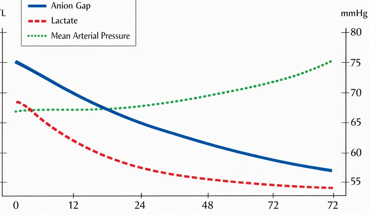
In septic shock, the anion gap and the lactate are followed together as markers of tissue perfusion and resuscitation adequacy. Serial values over the first 24 hours:
| Time | Na⁺ | Cl⁻ | HCO₃⁻ | AG | Lactate | MAP |
|---|---|---|---|---|---|---|
| 0 h (presentation) | 136 | 100 | 12 | 24 | 7.2 | 52 |
| 3 h (post-bolus) | 138 | 103 | 14 | 21 | 5.1 | 66 |
| 6 h | 139 | 104 | 16 | 19 | 3.6 | 71 |
| 12 h | 140 | 105 | 19 | 16 | 2.4 | 74 |
| 24 h | 139 | 106 | 22 | 11 | 1.6 | 78 |
The anion gap and lactate fall in parallel as mean arterial pressure (MAP) recovers with fluids, norepinephrine, and source control (the urine culture grew E. coli, sensitive to the empiric antibiotic). By 24 hours the gap has normalized (11 mEq/L), lactate is near-normal (1.6 mmol/L), and the bicarbonate has recovered to 22. The chloride has climbed to 106 — a mild hyperchloremic component from the large-volume saline resuscitation, which itself can narrow the gap slightly. The principles of serial anion gap and unmeasured-anion tracking in critically ill patients are covered in detail on the anion gap in the ICU page.
Key lessons from this case
- Lactate is the dominant unmeasured anion in septic shock — a high anion gap with an elevated lactate in a hypotensive patient is Type A lactic acidosis until proven otherwise.
- Always apply the albumin correction in septic patients, who are almost universally hypoalbuminemic; the corrected gap reveals the true magnitude of the acidosis.
- Lactate rarely explains the entire gap — concurrent AKI, stress ketogenesis, and phosphate accumulation contribute. Do not expect a perfect 1:1 lactate-to-gap correlation.
- Track the gap and lactate serially — both are perfusion surrogates, and their clearance predicts mortality in sepsis5. A persistently elevated gap after resuscitation prompts a search for ongoing hypoperfusion (e.g. mesenteric ischemia) or a second process.
References
- Kraut JA, Madias NE. "Serum Anion Gap: Its Uses and Limitations in Clinical Medicine." Clin J Am Soc Nephrol. 2007;2(1):162-174. Link ↗
- Figge J, et al. "The role of serum proteins in acid-base equilibria." J Lab Clin Med. 1998;117(6):453-467 (Figge albumin-corrected anion gap equation: AG + 2.5 × (4.0 − albumin g/dL)). See StatPearls — Biochemistry, Anion Gap. Link ↗
- Kharsa A, Edwards S. "Anion Gap and Non-Anion Gap Metabolic Acidosis." StatPearls [Internet]. Treasure Point (FL): StatPearls Publishing. Link ↗
- Life in the Fast Lane (LITFL). "Anion Gap." CCC Acid-Base. Link ↗
- Evans L, et al. "Surviving Sepsis Campaign: International Guidelines for Management of Sepsis and Septic Shock." Intensive Care Med. 2021;47(11):1181-1447 (lactate clearance as resuscitation target). See also MDCalc — Anion Gap. Link ↗